LATEST TECHNOLOGIES
The technology at Northern Beaches Cancer Clinic is cutting edge because your life depends on it. During your skin cancer consultation, you will likely encounter several advanced technologies.
At Northern Beaches Skin Cancer Clinic, we use advanced technology to integrate every step of analysis, recording, and clinical follow-ups for our patients. Our total body mapping and clinical dermoscopic imaging provide a concise and comprehensive clinical record that is unmatched.
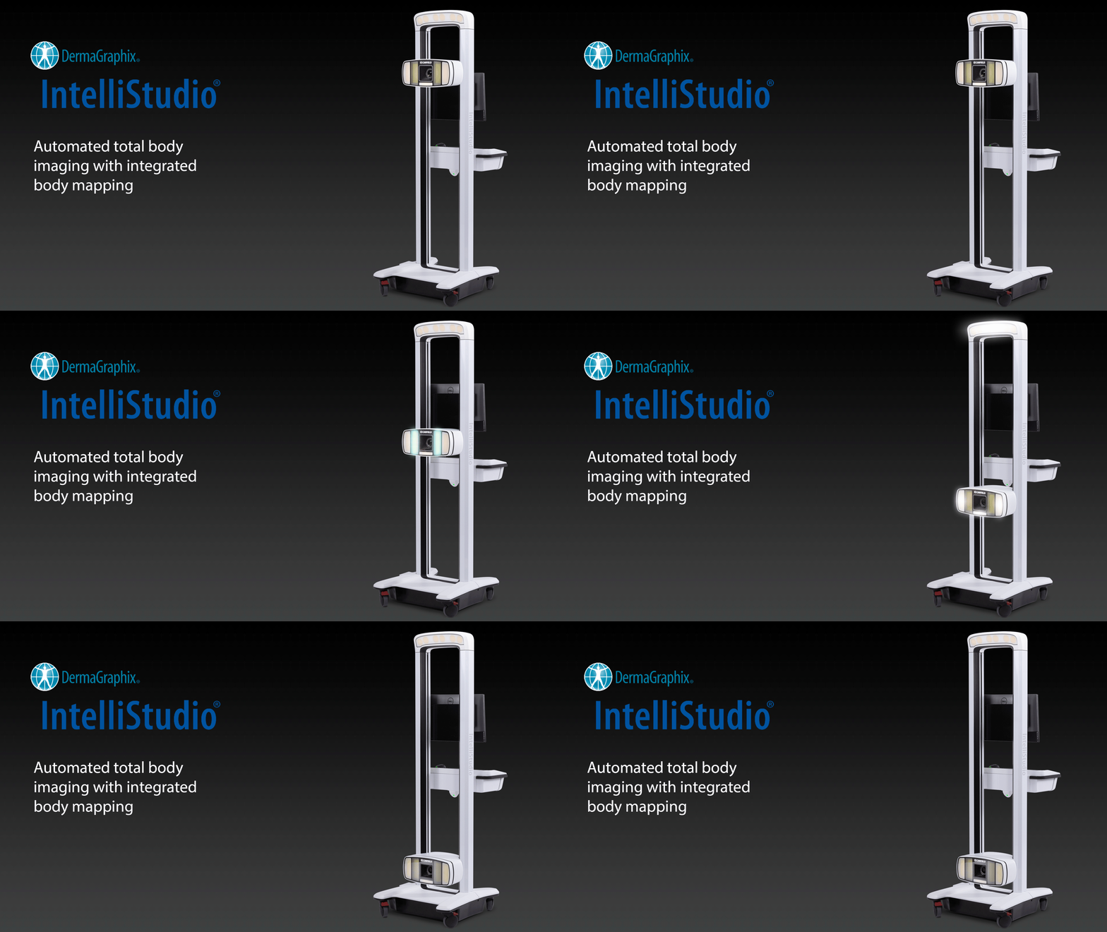
IntelliStudio®
Automated total body imaging with integrated body mapping
The Canfield IntelliStudio® is a state-of-the-art imaging system designed for comprehensive full-body photography. It is used to map and image the entire body, playing a crucial role in the early detection of melanoma. Equipped with a fully automated 50MP camera and Xenon smart dual capturing technology (white and cross-polarised light), IntelliStudio® ensures consistent, studio-quality images every time. By automating the process, it provides reliable, repeatable images that help medical professionals identify and monitor skin changes over time, aiding in the early diagnosis and treatment of melanoma.
DermLite DL3 & DL4
The DL3/4 have been developed to be the instrument of choice for the world’s leading skin cancer experts. Precision-engineered and crafted from solid aluminium, it is the first handheld DermLite to integrate a 25mm four-element lens, which offers greatly reduced optical distortion and a sharper image across the field of view.
Twenty-one high-powered LEDs produce approximately 30% more illumination in cross-polarized mode to create a brighter image. In other words, it means that your Dr can see all your spots more closely and monitor any changes that happen more easily.
Canfield d120
The professional image system for early recognition of skin cancer and image documentation with up to 120-fold magnification and HDplus image quality.
Canfield D120 is essential for progressive early recognition of skin cancer and image documentation. The examined skin lesion is directly displayed on the screen and can even be shown in different magnifications between 15 and 120 fold at the touch of a button on the camera.
Microprocessors, integrated in the Canfield D120 camera, adjust illumination intensity and colour mixture, so that there is a constant image quality for the follow-ups with your Dr over the years – it’s a very unique feature.
High quality standardized images are an indispensable requirement for reliable diagnosis.
Furthermore, clinical images of large skin areas can be made realistically by use of the macro function and can be printed in best photographic quality in order to document successful treatment.
The Canfield imaging system was developed within the course of the worldwide largest study for computer-assisted early recognition of skin cancer -“DANAOS“. After the image has been captured the integrated DANAOS system can calculate a classification value based on the world’s largest multi-centre study for computer-aided early detection of skin cancer. Borderline lesions can then be classified reliably and quickly by their dermoscopical relevant features using the ABCD rule.
Due to a close cooperation between well-known clinics and established doctors worldwide a technologically convincing system with a scientifically substantiated diagnostic support was created.
Decorated with a number of scientific and design awards, Canfield sets internationally new benchmarks in the field of computer-assisted early recognition of skin cancer.


